Overview
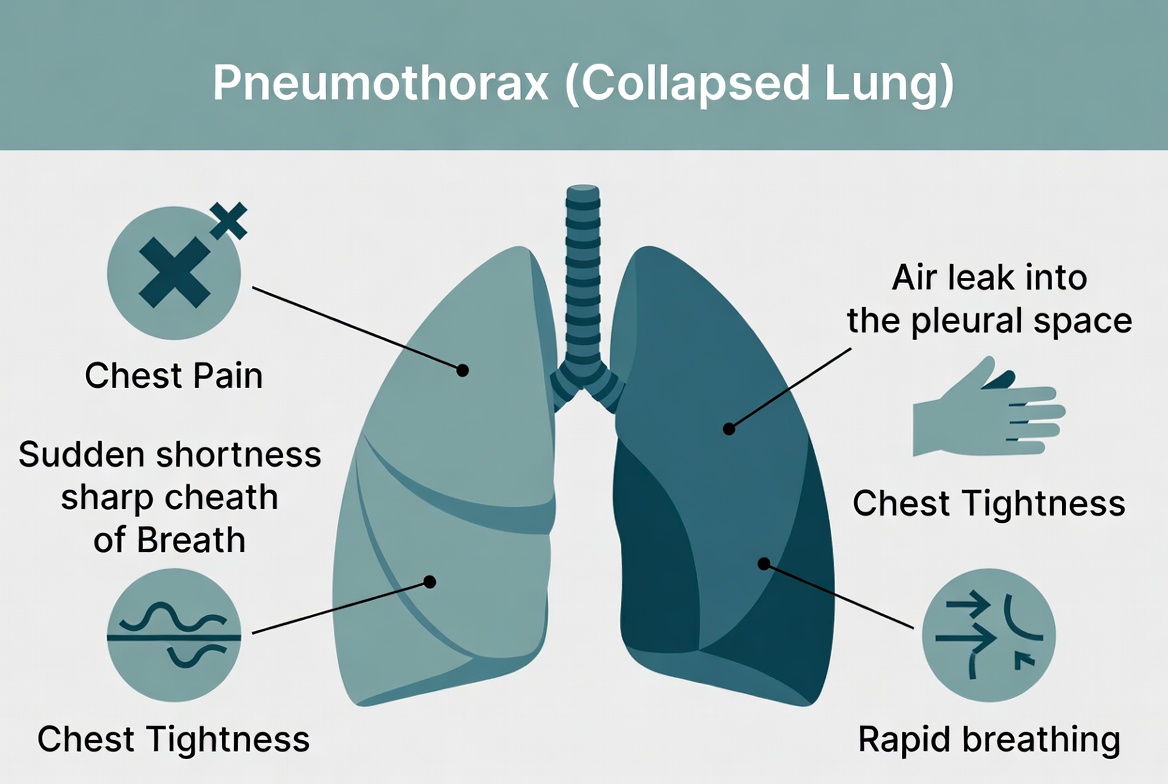
Pneumothorax, commonly known as a collapsed lung, occurs when air leaks into the space between the lung and the chest wall (the pleural space). This trapped air places pressure on the lung, causing it to partially or completely collapse and making it difficult to breathe.
A pneumothorax can occur spontaneously without an obvious cause, particularly in tall, thin young adults, or it may result from chest injury, underlying lung disease, or certain medical procedures. The severity ranges from a small pneumothorax that resolves on its own to a life-threatening tension pneumothorax that requires immediate emergency treatment.
Prompt diagnosis and appropriate treatment are essential to restore normal lung function, relieve symptoms, and prevent complications. Most people recover completely with timely medical care, although some may experience recurrent episodes.
Common Symptoms
- Sudden, sharp chest pain.
- Sudden shortness of breath.
- Chest tightness.
- Rapid breathing (tachypnea).
- Rapid heartbeat (tachycardia).
- Dry cough.
- Fatigue or reduced ability to exercise.
- Bluish lips or fingertips (cyanosis) in severe cases.
Tension Pneumothorax Is a Medical Emergency
A tension pneumothorax can rapidly become life-threatening by severely reducing blood flow to the heart and preventing normal lung expansion. Immediate emergency treatment is required.
Smoking Greatly Increases the Risk
Cigarette smoking significantly increases the risk of spontaneous pneumothorax, especially in young adults and people with underlying lung diseases such as COPD.
Early Treatment Leads to Excellent Recovery
Most people recover completely when pneumothorax is diagnosed early and treated appropriately. Following medical advice can also reduce the risk of recurrence.
If you develop sudden chest pain and shortness of breath, seek immediate medical evaluation. These symptoms should never be ignored, as they may indicate a pneumothorax or another serious medical emergency.
Symptoms
The symptoms of a pneumothorax (collapsed lung) usually begin suddenly and can range from mild to severe depending on the size of the air leak and the amount of lung collapse. Small pneumothoraces may cause only mild discomfort, while larger or tension pneumothoraces can rapidly become life-threatening.
Some people develop symptoms after a chest injury or medical procedure, while others experience a spontaneous pneumothorax without any obvious cause. People with underlying lung diseases such as chronic obstructive pulmonary disease (COPD) may have more severe symptoms even with a small pneumothorax.
Common Symptoms of Pneumothorax
| Symptom | Description |
|---|---|
| Sudden Chest Pain | The most common symptom. The pain is usually sharp, stabbing, and occurs on one side of the chest. |
| Sudden Shortness of Breath | Breathing becomes difficult because part of the lung has collapsed. |
| Chest Tightness | Some people feel pressure or tightness in the affected side of the chest. |
| Rapid Breathing (Tachypnea) | The body compensates for reduced lung function by increasing the breathing rate. |
| Rapid Heartbeat (Tachycardia) | The heart beats faster to help maintain oxygen delivery throughout the body. |
| Dry Cough | A persistent, non-productive cough may occur in some individuals. |
| Fatigue | Reduced oxygen levels and increased effort required for breathing can cause tiredness. |
| Bluish Lips or Fingertips (Cyanosis) | Low oxygen levels may cause a bluish discoloration, particularly in severe cases. |
Symptoms of a Tension Pneumothorax
A tension pneumothorax is a life-threatening emergency in which pressure continues to build inside the chest, compressing the lung, heart, and major blood vessels.
- Severe or rapidly worsening shortness of breath.
- Severe chest pain.
- Extreme difficulty breathing.
- Rapid heartbeat.
- Low blood pressure.
- Confusion or restlessness.
- Blue lips or fingertips (cyanosis).
- Loss of consciousness in severe cases.
Symptoms Usually Start Suddenly
Unlike many chronic lung diseases, pneumothorax typically causes sudden chest pain and shortness of breath that begin without warning. Symptoms should always be evaluated promptly.
People with Lung Disease May Have More Severe Symptoms
Individuals with COPD, asthma, cystic fibrosis, pulmonary fibrosis, or other chronic lung conditions may experience more severe breathing problems even with a relatively small pneumothorax.
Seek Emergency Medical Care Immediately If You:
- Develop sudden chest pain and shortness of breath.
- Experience severe or worsening difficulty breathing.
- Notice blue lips or fingertips.
- Feel faint, dizzy, or lose consciousness.
- Develop severe chest pain after a chest injury or medical procedure.
- Have symptoms that rapidly worsen over minutes.
A pneumothorax is often treatable, but delaying medical care can lead to serious complications. If you experience sudden chest pain or difficulty breathing, seek emergency medical evaluation immediately.
Causes
Pneumothorax (collapsed lung) occurs when air enters the pleural space—the area between the lung and the chest wall. As air accumulates in this space, pressure builds up around the lung, causing it to partially or completely collapse.
A pneumothorax may occur without an obvious cause (spontaneous), result from an injury to the chest (traumatic), develop because of an underlying lung disease (secondary spontaneous), or occur as a complication of certain medical procedures.
Common Causes of Pneumothorax
| Cause | Description |
|---|---|
| Primary Spontaneous Pneumothorax | Occurs without a known lung disease, often due to the rupture of small air-filled sacs (blebs) on the surface of the lung. It is most common in tall, thin young adults. |
| Secondary Spontaneous Pneumothorax | Develops as a complication of existing lung diseases such as chronic obstructive pulmonary disease (COPD), asthma, pulmonary fibrosis, cystic fibrosis, lung infections, or lung cancer. |
| Chest Trauma | Blunt injuries, rib fractures, stab wounds, gunshot wounds, or other chest injuries can allow air to leak into the pleural space. |
| Medical Procedures (Iatrogenic Pneumothorax) | Procedures such as lung biopsy, central venous catheter insertion, mechanical ventilation, thoracentesis, or chest surgery can occasionally cause a pneumothorax. |
| Mechanical Ventilation | High-pressure ventilation used in critically ill patients may cause overexpansion of the lungs and air leakage. |
| Ruptured Lung Blebs or Bullae | Fragile air sacs near the lung surface can burst spontaneously, allowing air to escape into the pleural space. |
| Rapid Changes in Air Pressure | Scuba diving, high-altitude climbing, or sudden changes in atmospheric pressure can increase the risk in susceptible individuals. |
| Rare Genetic or Connective Tissue Disorders | Conditions such as Marfan syndrome, Ehlers-Danlos syndrome, Birt-Hogg-Dubé syndrome, and alpha-1 antitrypsin deficiency may increase the risk of spontaneous pneumothorax. |
How Does a Pneumothorax Occur?
- Air leaks from the lung or enters through a chest injury.
- The air collects in the pleural space between the lung and chest wall.
- Pressure builds around the affected lung.
- The lung partially or completely collapses.
- If pressure continues to increase, a life-threatening tension pneumothorax may develop.
Spontaneous Pneumothorax Can Occur Without Warning
Many people with a primary spontaneous pneumothorax have no previous history of lung disease. The condition often develops suddenly after a small lung bleb ruptures.
Underlying Lung Disease Increases the Risk
People with COPD, pulmonary fibrosis, cystic fibrosis, severe asthma, lung infections, or lung cancer are more likely to develop a secondary spontaneous pneumothorax and often experience more severe symptoms.
Tension Pneumothorax Is a Medical Emergency
If air continues to build up in the pleural space, pressure can compress the lungs, heart, and major blood vessels. Immediate emergency treatment is required to prevent life-threatening complications.
Understanding the cause of a pneumothorax helps guide treatment and reduce the risk of recurrence. If you experience sudden chest pain or shortness of breath, seek immediate medical evaluation.
Risk Factors
Several factors can increase the likelihood of developing a pneumothorax (collapsed lung). Some people are more prone to spontaneous pneumothorax because of their body type or genetics, while others are at higher risk due to underlying lung diseases, smoking, chest injuries, or certain medical procedures.
Having one or more risk factors does not necessarily mean you will develop a pneumothorax, but understanding these factors can help you reduce your risk and recognize symptoms early.
Common Risk Factors for Pneumothorax
| Risk Factor | How It Increases Risk |
|---|---|
| Smoking | Smoking damages lung tissue and greatly increases the risk of spontaneous pneumothorax. The risk rises with the number of cigarettes smoked. |
| Tall, Thin Body Build | Primary spontaneous pneumothorax occurs more often in tall, thin adolescents and young adults, particularly males. |
| Male Sex | Men are more likely than women to develop primary spontaneous pneumothorax. |
| Age (15–34 Years) | Primary spontaneous pneumothorax is most common in teenagers and young adults, although pneumothorax can occur at any age. |
| Chronic Lung Diseases | Conditions such as COPD, asthma, cystic fibrosis, pulmonary fibrosis, tuberculosis, pneumonia, and lung cancer weaken the lungs and increase the risk of air leaks. |
| Previous Pneumothorax | People who have experienced a pneumothorax have a higher chance of recurrence, particularly during the first few years. |
| Chest Injury | Blunt trauma, rib fractures, stab wounds, gunshot wounds, and other chest injuries can lead to a collapsed lung. |
| Medical Procedures | Lung biopsy, thoracentesis, central venous catheter placement, chest surgery, and mechanical ventilation can occasionally cause pneumothorax. |
| Mechanical Ventilation | High airway pressures used during mechanical ventilation can increase the risk of lung rupture, particularly in critically ill patients. |
| Scuba Diving or High-Altitude Activities | Rapid changes in air pressure can increase the risk of lung injury and pneumothorax in susceptible individuals. |
| Genetic or Connective Tissue Disorders | Conditions such as Marfan syndrome, Ehlers-Danlos syndrome, Birt-Hogg-Dubé syndrome, and alpha-1 antitrypsin deficiency increase susceptibility to spontaneous pneumothorax. |
People at Higher Risk
- People who smoke.
- Tall, thin young adults, especially males.
- Individuals with COPD or other chronic lung diseases.
- People with a previous pneumothorax.
- Patients recovering from chest surgery or invasive medical procedures.
- People receiving mechanical ventilation.
- Scuba divers and high-altitude climbers.
- Individuals with inherited connective tissue or genetic disorders affecting the lungs.
Smoking Is the Most Preventable Risk Factor
Quitting smoking significantly lowers the risk of spontaneous pneumothorax and reduces the chance of recurrence after treatment.
Recurrence Is Common
After one episode of spontaneous pneumothorax, the risk of another episode is higher. Your healthcare provider may recommend follow-up care or procedures to reduce the likelihood of recurrence.
Managing Lung Disease Helps Reduce Risk
Controlling conditions such as COPD, asthma, cystic fibrosis, and pulmonary fibrosis, avoiding smoking, and following medical advice can help reduce the risk of developing a pneumothorax.
If you have multiple risk factors for pneumothorax or have experienced a previous collapsed lung, discuss prevention and follow-up care with your healthcare provider. Prompt evaluation of sudden chest pain or shortness of breath is essential for early diagnosis and treatment.
Diagnosis
Pneumothorax (collapsed lung) is diagnosed through a combination of medical history, physical examination, and imaging tests. Because its symptoms—such as sudden chest pain and shortness of breath—can resemble those of other heart and lung conditions, prompt evaluation is essential to confirm the diagnosis and determine the severity of the lung collapse.
The goals of diagnosis are to identify the presence of air in the pleural space, determine the size and type of pneumothorax, identify any underlying cause, and decide on the most appropriate treatment.
Common Tests Used to Diagnose Pneumothorax
| Test | Purpose |
|---|---|
| Medical History and Physical Examination | Your healthcare provider will ask about symptoms, recent chest injuries, smoking history, lung disease, recent medical procedures, and listen for reduced or absent breath sounds on the affected side. |
| Chest X-ray | The most common imaging test used to confirm a pneumothorax. It shows the presence and size of the collapsed lung and the amount of air in the pleural space. |
| Chest CT Scan | Provides detailed images of the lungs and pleural space. It can detect small pneumothoraces, ruptured blebs or bullae, underlying lung disease, and associated chest injuries. |
| Lung Ultrasound | A rapid bedside imaging test frequently used in emergency departments, trauma settings, and intensive care units to detect pneumothorax. |
| Pulse Oximetry | Measures blood oxygen saturation (SpO?) to assess how well oxygen is being delivered throughout the body. |
| Arterial Blood Gas (ABG) | Measures oxygen and carbon dioxide levels in the blood, especially in patients with severe breathing difficulty or chronic lung disease. |
| Electrocardiogram (ECG) | Helps rule out heart conditions such as a heart attack that can cause similar symptoms. |
| Blood Tests | Routine blood tests may be performed to evaluate overall health, identify infection, or prepare for procedures, although they do not diagnose pneumothorax directly. |
How Doctors Assess Severity
- Size of the pneumothorax.
- Amount of lung collapse.
- Severity of breathing difficulty.
- Blood oxygen levels.
- Presence of underlying lung disease.
- Signs of tension pneumothorax, such as low blood pressure, severe respiratory distress, or unstable vital signs.
Chest X-ray Is Usually the First Diagnostic Test
Most cases of pneumothorax are confirmed with a chest X-ray. If the diagnosis remains uncertain or more detailed information is needed, a chest CT scan or lung ultrasound may be performed.
Tension Pneumothorax Requires Immediate Treatment
If a tension pneumothorax is suspected based on symptoms and physical examination, emergency treatment may begin immediately without waiting for imaging, as delaying treatment can be life-threatening.
Early Diagnosis Improves Recovery
Prompt diagnosis allows healthcare providers to treat the collapsed lung before serious complications develop, improving recovery and reducing the risk of permanent lung damage.
If you experience sudden chest pain, shortness of breath, or symptoms following a chest injury or medical procedure, seek immediate medical evaluation. Early diagnosis and treatment are essential for the best possible outcome.
Treatment
Treatment for pneumothorax (collapsed lung) depends on the size of the air leak, the severity of symptoms, the type of pneumothorax, and the patient's overall health. The primary goals are to remove the trapped air, allow the lung to fully re-expand, relieve symptoms, and prevent future episodes.
Small pneumothoraces may heal on their own with careful monitoring, while larger or life-threatening cases require emergency procedures to remove the air from the pleural space.
Common Treatment Options for Pneumothorax
| Treatment | Purpose |
|---|---|
| Observation | Small pneumothoraces with mild symptoms may resolve naturally. Patients are monitored with repeat chest imaging to ensure the lung is re-expanding. |
| Supplemental Oxygen | Oxygen therapy improves blood oxygen levels and can help speed the absorption of air from the pleural space. |
| Needle Aspiration | A needle or small catheter is used to remove air from the pleural space, allowing the lung to re-expand. This is commonly used for selected stable patients with a primary spontaneous pneumothorax. |
| Chest Tube (Tube Thoracostomy) | A chest tube is inserted through the chest wall to continuously remove air until the lung fully re-expands. This is a common treatment for larger pneumothoraces. |
| Emergency Needle Decompression | For a tension pneumothorax, emergency needle decompression is performed immediately to relieve life-threatening pressure before placement of a chest tube. |
| Surgery | Video-assisted thoracoscopic surgery (VATS) or, less commonly, open surgery may be recommended for recurrent pneumothorax, persistent air leaks, or bilateral pneumothoraces. |
| Pleurodesis | A medical or surgical procedure that seals the space between the lung and chest wall to reduce the risk of future pneumothoraces. |
| Treatment of Underlying Lung Disease | Managing conditions such as COPD, asthma, pulmonary fibrosis, cystic fibrosis, or lung infections helps reduce recurrence and improve lung function. |
Recovery and Home Care
- Attend all follow-up appointments and chest imaging as recommended.
- Avoid smoking to reduce the risk of recurrence.
- Limit strenuous physical activity until your healthcare provider says it is safe.
- Avoid scuba diving unless cleared by a specialist, as it can significantly increase the risk of recurrence.
- Discuss the appropriate timing for air travel with your healthcare provider, as flying too soon after a pneumothorax may be unsafe.
- Take prescribed medications exactly as directed.
- Seek immediate medical care if symptoms return.
Many Small Pneumothoraces Heal Without Surgery
Small pneumothoraces often resolve with observation and oxygen therapy, but careful follow-up is essential to confirm that the lung has completely re-expanded.
Recurrent Pneumothorax May Require Surgery
People who experience repeated episodes or persistent air leaks may benefit from video-assisted thoracoscopic surgery (VATS) and pleurodesis to reduce the likelihood of future lung collapse.
Tension Pneumothorax Requires Immediate Emergency Treatment
A tension pneumothorax can rapidly become fatal if untreated. Emergency needle decompression followed by chest tube placement is the standard lifesaving treatment.
With prompt diagnosis and appropriate treatment, most people recover fully from a pneumothorax. Following your healthcare provider's recommendations and addressing risk factors such as smoking can significantly reduce the chance of recurrence.
Need Expert Medical Guidance?
Schedule a direct consultation with Dr. Nabila Zaheer for a personalized care plan.
Prevention
Not all cases of pneumothorax (collapsed lung) can be prevented, especially primary spontaneous pneumothorax. However, reducing modifiable risk factors and properly managing underlying lung conditions can significantly lower the risk of developing a pneumothorax or experiencing a recurrence.
People who have had a previous pneumothorax or have chronic lung disease should work closely with their healthcare provider to develop a personalized prevention plan.
Ways to Help Prevent Pneumothorax
| Prevention Strategy | How It Helps |
|---|---|
| Quit Smoking | Smoking is the most important preventable risk factor for spontaneous pneumothorax. Stopping smoking greatly reduces the risk of both first-time and recurrent episodes. |
| Manage Chronic Lung Diseases | Proper treatment of COPD, asthma, cystic fibrosis, pulmonary fibrosis, tuberculosis, and other lung conditions helps reduce lung damage and the likelihood of air leaks. |
| Attend Follow-Up Appointments | Regular medical reviews and follow-up chest imaging help confirm complete healing and detect recurrence early. |
| Avoid Scuba Diving After a Pneumothorax | Scuba diving can cause dangerous pressure changes that greatly increase the risk of another collapsed lung unless a specialist confirms it is safe. |
| Discuss Air Travel with Your Healthcare Provider | Flying before the lung has fully healed may worsen a pneumothorax due to changes in cabin pressure. Medical clearance is recommended after recovery. |
| Follow Safety Measures to Prevent Chest Injuries | Wearing seat belts, using protective equipment during sports, and following workplace safety practices reduce the risk of traumatic pneumothorax. |
| Follow Medical Instructions After Procedures | After chest surgery or procedures involving the lungs, following post-procedure instructions helps reduce complications and supports proper healing. |
| Consider Preventive Surgery if Recommended | People with recurrent pneumothoraces or persistent air leaks may benefit from procedures such as pleurodesis or video-assisted thoracoscopic surgery (VATS) to reduce recurrence. |
Tips for Reducing the Risk of Recurrence
- Do not smoke or use tobacco products.
- Take medications for chronic lung disease exactly as prescribed.
- Seek prompt treatment for respiratory infections.
- Avoid activities involving major pressure changes unless medically cleared.
- Know the warning signs of recurrence, including sudden chest pain and shortness of breath.
- Maintain regular follow-up with your healthcare provider if you have had a previous pneumothorax.
Smoking Cessation Makes a Significant Difference
People who stop smoking after a pneumothorax have a substantially lower risk of experiencing another collapsed lung compared with those who continue to smoke.
Be Cautious with Flying and Diving
Do not fly or scuba dive until your healthcare provider confirms that your lung has completely healed. Pressure changes during these activities can trigger or worsen a pneumothorax.
Healthy Lungs Reduce Future Risk
Managing chronic lung diseases, avoiding smoking, preventing chest injuries, and following medical advice after treatment can help reduce the likelihood of future pneumothoraces and support long-term lung health.
Although a pneumothorax cannot always be prevented, recognizing your risk factors and taking preventive measures can lower the chance of recurrence. If you develop sudden chest pain or shortness of breath, seek immediate medical attention.
Complications
Although many cases of pneumothorax (collapsed lung) resolve completely with prompt treatment, complications can occur if the condition is severe, treatment is delayed, or underlying lung disease is present. Large pneumothoraces and tension pneumothoraces are particularly dangerous and require immediate medical attention.
Early diagnosis and appropriate treatment greatly reduce the risk of serious complications and improve long-term outcomes.
Possible Complications of Pneumothorax
| Complication | Description |
|---|---|
| Tension Pneumothorax | Air continues to build up in the pleural space, creating pressure that compresses the lung, heart, and major blood vessels. This is a life-threatening medical emergency. |
| Respiratory Failure | Severe lung collapse can prevent adequate oxygen from reaching the body, resulting in respiratory failure, particularly in people with underlying lung disease. |
| Low Blood Oxygen Levels (Hypoxemia) | A collapsed lung reduces oxygen exchange, which may lead to dangerously low oxygen levels in the bloodstream. |
| Recurrent Pneumothorax | Some people experience repeated episodes, especially if they continue smoking or have persistent lung abnormalities such as blebs or bullae. |
| Persistent Air Leak | Air may continue leaking from the lung for several days despite treatment, sometimes requiring surgery or pleurodesis. |
| Infection | Although uncommon, infection can develop around a chest tube or after surgical treatment if bacteria enter the pleural space. |
| Bleeding (Hemothorax) | Chest trauma or certain procedures may cause bleeding into the pleural space along with pneumothorax. |
| Lung Scarring (Pleural Thickening) | Healing after recurrent pneumothorax or surgery may lead to scar tissue that affects the pleura. |
| Cardiovascular Collapse | Untreated tension pneumothorax can severely reduce blood return to the heart, causing shock, cardiac arrest, and death. |
Warning Signs of Complications
- Sudden worsening of shortness of breath.
- Severe or increasing chest pain.
- Blue lips or fingertips (cyanosis).
- Rapid heartbeat or very low blood pressure.
- Dizziness, fainting, or confusion.
- Persistent air leak from a chest tube.
- Fever, redness, swelling, or drainage around a chest tube insertion site.
- Return of symptoms after treatment.
Tension Pneumothorax Can Be Fatal
Without immediate treatment, a tension pneumothorax can rapidly lead to respiratory failure, cardiovascular collapse, cardiac arrest, and death. Seek emergency medical care immediately if symptoms suddenly worsen.
Recurrence Is Possible
People who have experienced one pneumothorax are at increased risk of another. Stopping smoking and following your healthcare provider's recommendations can significantly reduce this risk.
Most People Recover Completely
With early diagnosis, appropriate treatment, and regular follow-up, most people recover fully from a pneumothorax and return to their normal daily activities.
If you develop worsening shortness of breath, severe chest pain, fainting, or symptoms that return after treatment, seek immediate medical attention. Prompt recognition of complications can be lifesaving and helps protect long-term lung function.
When to See a Doctor
A pneumothorax (collapsed lung) can range from a small air leak that causes mild symptoms to a life-threatening emergency. Because symptoms often begin suddenly and may worsen quickly, it is important to seek medical evaluation as soon as they occur.
Early diagnosis and treatment help prevent complications such as respiratory failure, tension pneumothorax, and cardiovascular collapse.
Schedule a Medical Appointment If You:
- Develop mild chest pain that persists or recurs.
- Notice new or unexplained shortness of breath.
- Have a dry cough that continues without improvement.
- Recently recovered from a pneumothorax and develop new respiratory symptoms.
- Have a chronic lung disease, such as COPD, asthma, cystic fibrosis, or pulmonary fibrosis, and experience worsening breathing.
- Recently underwent a chest procedure or surgery and notice chest discomfort or increasing breathlessness.
- Have a history of spontaneous pneumothorax and want guidance before flying or scuba diving.
Seek Emergency Medical Care Immediately If You:
- Develop sudden, severe chest pain.
- Experience sudden or worsening shortness of breath.
- Have difficulty breathing that rapidly gets worse.
- Notice blue lips or fingertips (cyanosis).
- Feel faint, dizzy, confused, or lose consciousness.
- Develop chest pain or breathing difficulty after a chest injury.
- Experience symptoms following a lung biopsy, central venous catheter placement, mechanical ventilation, or another chest procedure.
- Have rapid heartbeat, low blood pressure, or signs of shock, which may indicate a tension pneumothorax.
Do Not Ignore Sudden Chest Pain
Sudden chest pain and shortness of breath are the most common symptoms of a pneumothorax. These symptoms can also occur with other serious conditions such as a heart attack or pulmonary embolism, so immediate medical evaluation is essential.
People with Previous Pneumothorax Should Stay Alert
If you have had a collapsed lung before, seek medical attention promptly if similar symptoms return, as recurrence is relatively common.
Prompt Treatment Improves Recovery
Most people recover completely when a pneumothorax is diagnosed and treated early. Timely medical care helps prevent complications and reduces the risk of permanent lung damage.
Never ignore sudden chest pain, shortness of breath, or breathing difficulty, especially after a chest injury or medical procedure. If symptoms are severe or worsen rapidly, call your local emergency services or go to the nearest emergency department immediately.